O Level Biology 34 Views 1 Answers
Sourav PanLv 9November 3, 2024
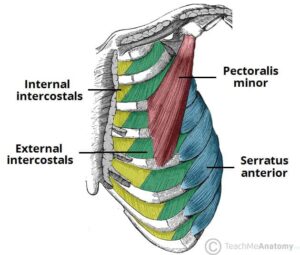
Identify, on diagrams and images, the ribs, internal and external intercostal muscles and the diaphragm
Identify, on diagrams and images, the ribs, internal and external intercostal muscles and the diaphragm
Please login to save the post
2
Please login to submit an answer.
Sourav PanLv 9May 15, 2025
To identify the components of the thoracic cavity, including the ribs, internal and external intercostal muscles, and diaphragm, here is a detailed description of each structure along with their roles in respiration:
Ribs
- Description: The ribs are a series of long, curved bones that form the rib cage. There are 12 pairs of ribs in humans, which protect the thoracic cavity and support the upper body.
- Role: They provide structural support for the thoracic cavity and play a crucial role in breathing by expanding and contracting during inhalation and exhalation.
External Intercostal Muscles
- Description: These muscles are located between the ribs and run from the lower border of one rib to the upper border of the rib below it. They are oriented inferomedially (downward and inward).
- Role: The external intercostal muscles are primarily involved in inhalation. When they contract, they elevate the ribs, increasing the volume of the thoracic cavity and allowing air to flow into the lungs.
Internal Intercostal Muscles
- Description: Situated deep to the external intercostals, these muscles run in the opposite direction (from the upper border of one rib to the lower border of the rib above). They have two parts: interosseous (between ribs) and interchondral (between costal cartilages).
- Role: The internal intercostal muscles assist in forced expiration by depressing the ribs and reducing the volume of the thoracic cavity, which helps push air out of the lungs.
Diaphragm
- Description: The diaphragm is a dome-shaped muscle that separates the thoracic cavity from the abdominal cavity. It is attached to the lower ribs, sternum, and lumbar vertebrae.
- Role: The diaphragm is the primary muscle of respiration. During inhalation, it contracts and flattens, increasing thoracic volume and drawing air into the lungs. During exhalation, it relaxes, reducing thoracic volume and expelling air.

0
0 likes
- Share on Facebook
- Share on Twitter
- Share on LinkedIn
0 found this helpful out of 0 votes
Helpful: 0%
Helpful: 0%
Was this page helpful?


