Taenia Solium
- Taenia solium, also known as the pork tapeworm, is a parasitic flatworm that infects humans and pigs. It belongs to the class Cestoda and is one of the most common human tapeworms worldwide.
- Taenia solium infection occurs when a person ingests undercooked or raw pork that contains the tapeworm larvae. The larvae then develop into adult tapeworms in the small intestine, where they can grow up to several meters in length. The adult tapeworms can produce thousands of eggs that are shed in the feces and can contaminate the environment.
- Humans can also become infected with Taenia solium by ingesting food or water contaminated with tapeworm eggs. Once ingested, the eggs hatch into larvae that can migrate to various organs in the body, including the brain, where they can cause a condition known as neurocysticercosis. This can lead to seizures, headaches, and other neurological symptoms.
- Prevention of Taenia solium infection involves proper cooking of pork and good hygiene practices, such as washing hands and properly disposing of human and animal waste. Treatment for Taenia solium infection usually involves the use of antiparasitic drugs, such as praziquantel, in combination with supportive care for any associated symptoms.
- Taenia solium infection is a significant public health concern, particularly in developing countries with poor sanitation and hygiene practices. Control measures include education on proper food handling and cooking practices, improved sanitation, and the use of anthelmintic drugs in both humans and animals.

Characteristics of Taenia Solium
Taenia solium, also known as the pork tapeworm, is a parasitic flatworm that infects humans and pigs. Here are some of its characteristics:
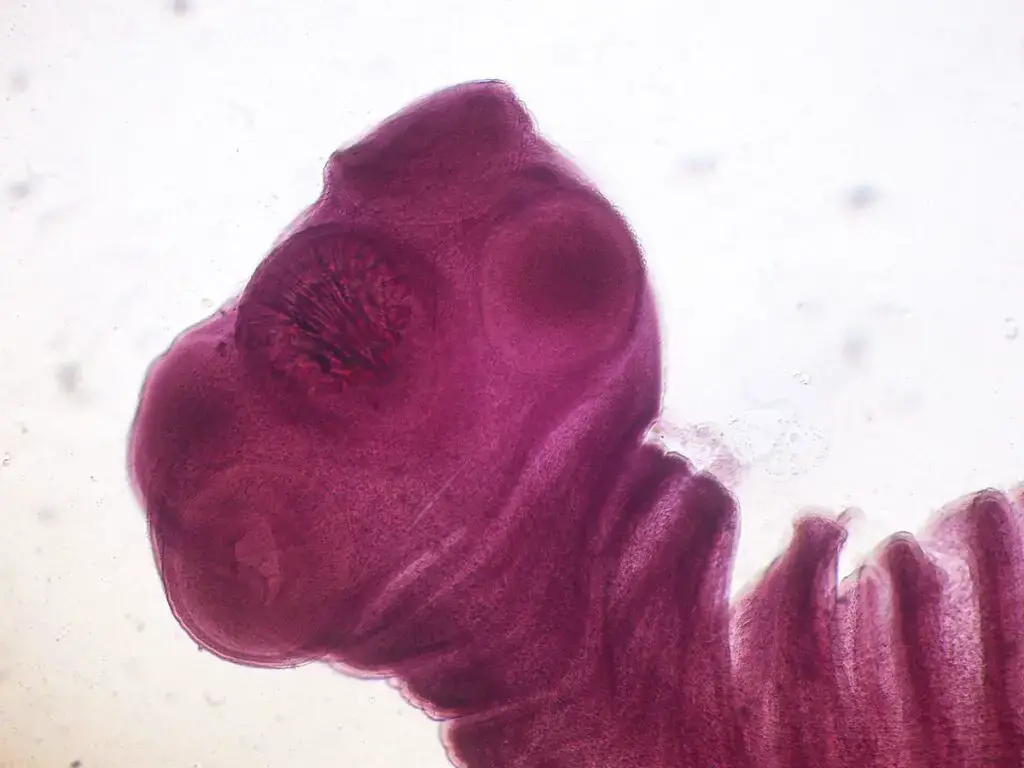
- Morphology: The adult Taenia solium tapeworm can grow up to several meters in length and has a long, flat body composed of multiple segments called proglottids. The head of the tapeworm, known as the scolex, has hooks and suckers that allow it to attach to the host’s intestinal wall.
- Life cycle: The life cycle of Taenia solium involves two hosts – pigs and humans. The adult tapeworms live in the small intestine of humans and produce eggs that are excreted in the feces. The eggs can contaminate soil, water, or food, and can be ingested by pigs. The eggs hatch into larvae in the pig’s intestines, which can then migrate to the pig’s muscles and form cysts. If humans eat undercooked pork containing cysts, the cysts can develop into adult tapeworms in the human intestine.
- Transmission: Taenia solium is transmitted to humans through ingestion of undercooked or raw pork containing cysts or through ingestion of food or water contaminated with tapeworm eggs. It is most commonly found in areas with poor sanitation and hygiene practices.
- Symptoms: Many people with Taenia solium infection do not experience any symptoms, but some may experience abdominal pain, nausea, diarrhea, or weight loss. In rare cases, the larvae can migrate to the brain and cause neurocysticercosis, which can lead to seizures, headaches, and other neurological symptoms.
- Diagnosis: Diagnosis of Taenia solium infection can be challenging, as the symptoms can be similar to other gastrointestinal diseases. Blood tests and stool exams can help detect the presence of the tapeworm or its eggs.
- Treatment: The treatment of Taenia solium infection involves the use of antiparasitic drugs, such as praziquantel or albendazole, to kill the adult tapeworms or larvae. Surgery may also be necessary in cases of neurocysticercosis.
- Prevention: The best way to prevent Taenia solium infection is by properly cooking pork to a temperature of at least 145°F (63°C) and practicing good hygiene, such as washing hands regularly and avoiding contact with feces or contaminated soil, water, or food.
Life history of Taenia solium
- While still inside the proglottid, fertilized eggs transform into six-hooked Heacanth larva.
- They then create cysts in the pig’s voluntary muscle after they’ve wriggled their way into the body.
- At this point, the cyst’s wall has developed into a distinct head, at which point it is referred to as Cysticercus.
- When the Cysticercus reaches the human population, it matures and begins to release a proglottid series.
Fasciola Hepatica
- Fasciola hepatica, also known as the common liver fluke, is a parasitic flatworm that infects the liver of various mammals, including humans, sheep, cattle, and deer. It belongs to the class Trematoda and is one of the most economically important helminth parasites of livestock worldwide.
- The adult fluke lives in the bile ducts of the host’s liver and feeds on blood, causing damage to the liver tissue and reducing the host’s productivity. The life cycle of Fasciola hepatica involves two intermediate hosts, a freshwater snail and a plant-eating mammal, such as a cow or sheep.
- Humans can become infected with Fasciola hepatica by ingesting contaminated water or vegetation, such as watercress or other freshwater plants. The disease caused by Fasciola hepatica is called fascioliasis and can cause symptoms such as fever, abdominal pain, and liver damage.
- Diagnosis of fascioliasis can be challenging, as the symptoms can be similar to other liver diseases, and the eggs produced by the fluke may not always be detectable in stool samples. However, blood tests and imaging studies such as ultrasound can help confirm the diagnosis.
- Treatment for fascioliasis usually involves the use of antiparasitic drugs such as triclabendazole, but prevention is also important. Measures to prevent infection include avoiding consumption of contaminated water and vegetation, avoiding contact with infected animals, and practicing good hygiene.
- Fasciola hepatica infection is a significant public health concern, particularly in areas where people rely on freshwater resources for food and water. Control measures include education on proper food and water handling practices, improved sanitation, and the use of anthelmintic drugs in both humans and animals.

Characteristics of Fasciola Hepatica
Fasciola hepatica is a parasitic flatworm that infects the liver and bile ducts of various mammals, including humans. Here are some of its characteristics:
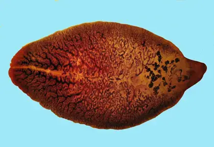
- Morphology: Adult Fasciola hepatica worms are flat and leaf-like in shape, with a length of about 20-30 mm and a width of 5 mm. They have a tapered anterior end and a broader posterior end. They have two suckers, one oral sucker at the anterior end and one ventral sucker at the mid-body region, which help them attach to the host’s intestinal wall.
- Life cycle: The life cycle of Fasciola hepatica involves two hosts – a snail intermediate host and a mammalian definitive host. The adult worms live in the bile ducts of the definitive host and produce eggs that are excreted in the feces. The eggs hatch into larvae in water, and the larvae infect snails, where they undergo several developmental stages before being released into water as infective cercariae. The cercariae can then penetrate the skin of the definitive host and migrate to the liver, where they develop into adult worms.
- Transmission: Fasciola hepatica is transmitted to humans through ingestion of contaminated water or vegetables, especially watercress, that contain metacercariae (encysted larvae) of the parasite. It is most commonly found in areas with poor sanitation and hygiene practices.
- Symptoms: Many people with Fasciola hepatica infection do not experience any symptoms, but some may experience abdominal pain, nausea, diarrhea, or jaundice. In chronic cases, the infection can lead to liver damage and fibrosis.
- Diagnosis: Diagnosis of Fasciola hepatica infection can be challenging, as the symptoms can be similar to other liver diseases. Blood tests and imaging studies, such as ultrasound or computed tomography (CT) scan, can help detect the presence of the parasite or its eggs.
- Treatment: The treatment of Fasciola hepatica infection involves the use of antiparasitic drugs, such as triclabendazole or albendazole, to kill the adult worms or larvae. Surgery may also be necessary in cases of severe liver damage.
- Prevention: The best way to prevent Fasciola hepatica infection is by avoiding ingestion of contaminated water or vegetables and by practicing good hygiene, such as washing hands regularly and properly cooking food.
Life history of Fasciola Hepatica
- The fertilized egg develops into a ciliated Miracidium larva that invades the body of the mollusk.
- Miracidium transforms into Sporocyst, where Rediae develop.
- Rediae produce daughter Rediae.
- Rediae (daughter) give rise to Cercariae, which, upon reaching their principal host (sheep), transform into adult flukes.
Differences Between Taenia Solium and Fasciola Hepatica
| Characteristics | Taenia Solium | Fasciola Hepatica |
|---|---|---|
| Body shape | Elongated, divided into a head, neck, and proglottides | Flat, unsegmented, and leaf-like |
| Head structure | Equipped with hooks and suckers | Has a bifid mouth and a pair of suckers (anterior and posterior) |
| Alimentary canal | Absent | Bifid alimentary canal with multiple caeca |
| Nervous system | Composed of two undifferentiated ganglia and nerve cords | Composed of two differentiated ganglia and nerve cords |
| Excretory system | Well-developed | More developed |
| Reproductive unit | Each proglottis is a self-sufficient reproductive unit with both male and female organs | Whole animal is a single reproductive unit |
| Fertilization | Self-fertilization between different segments of the same individual or between different individuals | Cross-fertilization between different individuals |
| Larval form | Feeble movement of contraction and expansion, no sense organ | Well-developed cilia as locomotory organelles (miracidium), presence of eye spots |
| Asexual reproduction | No asexual method of larval reproduction | Asexual method of larval reproduction present (rediae) |
| Larval life | Less complicated | More complicated |
| Strobilation | Less risk of loss or death | More risky, especially as the presence of water is essential |
| Life cycle | Completed in two hosts that belong to the class Mammalia | Completed in two hosts, one belonging to the vertebrate and the other to the invertebrate. |
| Feature | Fasciola Hepatica | Taenia Solium |
|---|---|---|
| Habitat | Bile duct of liver of sheep, cows, pigs, etc. (occasionally found in humans) | Alimentary canal of humans (cyst forms occur in pigs) |
| Geographical distribution | Asia, America, Europe, Africa | Worldwide |
| Body shape | Flat and leaf-like | Flat and ribbon-like |
| Size | 1.0-2.5 cm in length, width about 1 cm | Up to 3 meters in length |
| Anterior end | Conically projected to form the ‘head lobe’ | Knob-like head or ‘scolex’ with double rows of curved chitinous hooks and four suckers |
| Suckers | Two (anterior and ventral) | Four |
| Genital pore | Single median, situated ventrally and in between the two suckers | Repeated in each proglottid |
| Excretory aperture | Single, situated at the extreme posterior end of the body | Ends of lateral canals act as excretory pores |
| Alimentary canal | Short oesophagus, bifurcates to form the intestine. Many caeca originate from the main branches almost in every region of the body. There is no anus. | Absent, absorption occurs through general body wall predominantly by diffusion |
| Excretory system | Main excretory canal opens to the exterior through an aperture situated at the posterior end of the body. The duct receives many tubules each of which ends in flame bulb. | Many flame-bulbs arranged superficially and having drainage tubules. These tubules carry the excretory products to two longitudinal canals placed laterally. |
| Nervous system | Consists of prominent masses, called cerebral ganglia, joined together by a nerve ring around the oesophagus. From the ganglia nerves are given off to the head lobe and to the posterior part of the body. One pair of these posterior nerves is larger and more stout than the others. | Scolex bears a transverse band of nervous material. This ganglionated mass gives out slender branches to the suckers and to the posterior parts of the body. Two stouter nerves run laterally along the long axis of the body. |
| Reproduction | Hermaphrodite. Sex organs are well-developed. The male organs consist of a pair of much-branched testes, two vasa deferentia, a pear-shaped seminal vesicle, a convoluted ejaculatory duct and a muscular penis (cirrus). Female organs consist of single and branched ovary, a convoluted oviduct, shell gland, a uterus, which leads to the genital pore. Accessory glands – yolk or vitelline glands – open through ducts in the shell gland. A canal called Laurer’s canal running between the point of fusion of vitelline duct and oviduct at one end and the dorsal surface at the other end. Cross-fertilization is the rule. | Hermaphrodite. Sex organs are repeated in each proglottid. A mature proglottid is almost filled with sex organs. Male organs consist of numerous testes, efferent ducts, vas deferens and a protrusible penis (cirrus) lying in the genital atrium located on one of the lateral borders. Female organs consist of ovary which is bilobed and situated at the hinder end – a short oviduct, and vagina which opens into the genital atrium. Uterus is much branched |
| Characteristic | Fasciola Hepatica | Taenia Solium |
|---|---|---|
| Life Cycle | Indirect, involves freshwater snail as intermediate host | Indirect, involves pigs as intermediate host |
| Infection in humans | Ingestion of metacercariae on vegetation or in contaminated water | Ingestion of undercooked infected pork |
| Geographic Distribution | Asia, America, Europe, Africa | Worldwide |
| Anatomy | Flat, leaf-like body with conically projected head lobe, two suckers, and a single median genital pore. No anus. | Flat, ribbon-like body consisting of a knob-like scolex with four suckers and double rows of curved chitinous hooks, and a chain of proglottids with numerous testes, a uterus, and a compact vitelline gland. No alimentary canal. |
| Size | 1.0-2.5 cm in length and 1 cm in width | Up to 3 meters in length, with proglottids increasing in size from anterior to posterior |
| Symptoms of infection | Abdominal pain, nausea, diarrhea, anemia, and liver damage | Usually asymptomatic, but may cause mild abdominal discomfort, diarrhea, and weight loss |
| Treatment | Praziquantel or triclabendazole | Praziquantel or niclosamide |
FAQ
What are Taenia Solium and Fasciola Hepatica?
Answer: Taenia Solium and Fasciola Hepatica are parasitic worms that affect humans and animals.
What is the difference in the habitat of Taenia Solium and Fasciola Hepatica?
Answer: Taenia Solium lives in the alimentary canal of humans, while Fasciola Hepatica lives in the bile duct of the liver of sheep, cows, pigs, etc. and occasionally in humans.
How do Taenia Solium and Fasciola Hepatica differ in their body shape?
Answer: Taenia Solium has a flat and ribbon-like body that consists of a knob-like head or ‘scolex’ and a great number of similar parts, called proglottids, arranged in a single row. On the other hand, Fasciola Hepatica has a flat and leaf-like body, size varies from 1.0 to 2.5 cm in length, and width is about 1 cm.
What is the difference between the mouth of Taenia Solium and Fasciola Hepatica?
Answer: Taenia Solium doesn’t have an alimentary canal and absorption occurs through the general body wall predominantly by diffusion. Fasciola Hepatica has a mouth that lies in the middle of the anterior sucker and is followed by a suctorial pharynx.
How do Taenia Solium and Fasciola Hepatica differ in their excretory system?
Answer: In Taenia Solium, many flame-bulbs are arranged superficially in each proglottid and have drainage tubules. These tubules carry the excretory products to two longitudinal canals placed laterally. On the other hand, the main excretory canal of Fasciola Hepatica opens to the exterior through an aperture situated at the posterior end of the body, and the duct receives many tubules each of which ends in a flame bulb.
How do Taenia Solium and Fasciola Hepatica differ in their nervous system?
Answer: Taenia Solium bears a transverse band of nervous material on the scolex, and this ganglionated mass gives out slender branches to the suckers and posterior parts of the body. In contrast, Fasciola Hepatica’s nervous system consists of prominent masses called cerebral ganglia, joined together by a nerve ring around the oesophagus, and from the ganglia, nerves are given off to the head lobe and the posterior part of the body.
What is the difference in the reproductive system of Taenia Solium and Fasciola Hepatica?
Answer: Both Taenia Solium and Fasciola Hepatica are hermaphrodite. However, in Taenia Solium, sex organs are repeated in each proglottid, while a mature proglottid is almost filled with sex organs. In Fasciola Hepatica, there is a single median genital pore situated ventrally and between the two suckers.
What is the difference in the size of Taenia Solium and Fasciola Hepatica?
Answer: Taenia Solium can grow up to 3 meters long and has a large number of proglottids, while Fasciola Hepatica’s size varies from 1.0 to 2.5 cm in length and has a leaf-like shape.
Do Taenia Solium and Fasciola Hepatica have different geographical distribution?
Answer: Yes, Taenia Solium is found worldwide, while Fasciola Hepatica is found in Asia, America, Europe, and Africa.
Are Taenia Solium and Fasciola Hepatica harmful to humans?
Answer: Yes, both Taenia Solium and Fasciola Hepatica are harmful to humans and can cause various health issues, including diarrhea, abdominal
- Text Highlighting: Select any text in the post content to highlight it
- Text Annotation: Select text and add comments with annotations
- Comment Management: Edit or delete your own comments
- Highlight Management: Remove your own highlights
How to use: Simply select any text in the post content above, and you'll see annotation options. Login here or create an account to get started.