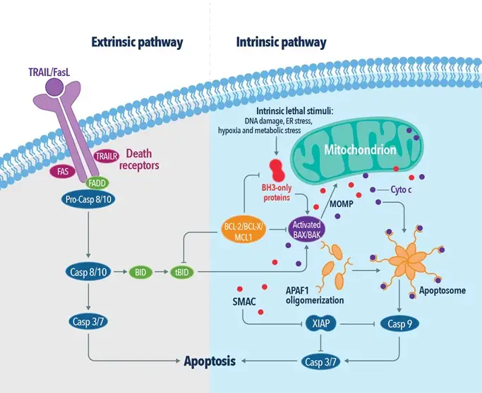
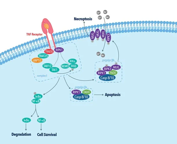
Multicellular organisms experience a variety of cell development and death processes. Cellular death is crucial for the survival and development of all organisms. Our body is made up of several cell types situated in diverse body sections. There are two distinct methods by which a cell dies. Either the organism is exposed to an unfavourable or dangerous environment, sustains an injury, or undergoes a controlled disintegration process. A cell dies by two distinct processes, called Apoptosis and Necrosis.
Cell death may be caused by a variety of hazardous chemicals or even physical events, such as radiation, trauma, heat, and loss of oxygen delivery owing to decreased blood flow. All of these chemical or physical occurrences can result in a fatal alteration of the cell’s structure and function.
Apoptosis and Necrosis are both forms of cell death that differ considerably in every areas except the conclusion. Necrosis is a type of cell death in which the cell dies prematurely as a result of uncontrolled external forces. Apoptosis is characterised as a predetermined suicide cell in which the cell destroys itself to maintain the body’s normal function.
What is Apoptotic and Necrotic Processes?

- Both apoptosis and necrosis can be viewed as components of a spectrum of related biochemical processes that both lead to cellular death in one way or another.
- Cells that have undergone apoptosis, also known as programmed cell death (PCD), shrink, develop blebs (bubble-like patches) on their membranes, have the genetic and protein content of their nuclei degraded, and have their mitochondrial structure break down, releasing cytochrome.
- Other molecules (such ATP and UTP) are readily released while the pieces are individually covered in their own membrane. These substances direct cell-eating organisms known as macrophages to seek for and consume dead cells and their fragments.
- A phospholipid that is ordinarily inert in a cell’s membrane causes this “eat me” communication to be sent, and the macrophages in response release cytokines that suppress inflammatory reactions.
- Necrotic cells, on the other hand, lose their chemical makeup and processes, swell or may form vacuoles on their surface, and their internal structures may expand or contract quickly.
- In addition to causing swelling (inflammation) and edoema in the surrounding tissues, the uncontrolled release of cytochrome and the phospholipid known as phosphatidylserine from the cell membrane frequently results in neighbouring cells dying through apoptosis.
- Necrotic cells, unlike those that have undergone apoptosis, are not targeted by macrophages for the removal of their cellular debris, allowing the effects of the cell rupture to spread fast and for a very long time throughout the body.
- The process of apoptosis is energy-dependent, which means that a cell must provide energy for it to proceed, giving rise to the term “cell suicide.” Necrosis is caused by external forces or localised illnesses, and necrosis does not require any energy input from a cell.
- The main chemical cues for the apoptotic pathways that result in cell demise are inactive proenzymes termed caspases. Because a cell is destroyed in an uncontrolled manner during necrotic processes, necrosis occasionally uses caspases but to a much smaller extent and frequently does not.
- Necrosis, for instance, is the process that results in the dead or necrotic tissue that surrounds, let’s say, a bite from a poisonous spider.
- There may be as many as 13 caspases, which have been roughly grouped as inflammatory, effectors or executioners (those that really cause cell death), and initiators. Contrary to popular belief, inflammatory caspases actually prevent inflammation. Since the inflammatory caspase input is absent during necrosis, inflammation is always present during necrotic cell death.
Symptoms of Apoptotic and Necrotic
- There are no outward signs of the process because apoptosis is a normal component of an organism’s cellular equilibrium. Necrosis, on the other hand, is an uncontrolled alteration of an organism’s cell balance, making it constantly dangerous and producing audible, unfavourable signs.
- As parts (such as cell structures, cytoplasm, and DNA/RNA) of the ruptured or damaged cells are liberated, necrosis is initially followed by inflammation.
- An organism’s response to this uncontrolled flow of proteins, chemicals, and genetic material includes inflammation to protect nearby tissues as well as an increase in the creation of white blood cells, macrophages, and T cells to combat infection.
- A metabolic surge and fever are frequent side effects of these reactions, which can cause exhaustion and an all-around compromised immune system.
- Necrotic tissues will lose their vascularity, or blood flow, if untreated, and begin to deteriorate. The condition where tissue eventually dies and needs to be removed to halt necrosis from spreading is called gangrene when this occurs.

Causes of Apoptosis and Necrosis
Cell death can be brought on by three different mechanisms:
- signals produced by a cell on its own, which may be brought on by ageing, infections, erroneous mitosis (cell division), or other factors. The following two types of cell death are extrinsic processes, whereas this mechanism is known as the intrinsic or mitochondrial pathway.
- the induction of death activators, cell surface receptors that react to outside cues like hormones or other chemical messengers.
- external stimulation by potentially harmful reactive oxygen species such free radicals.
Apoptosis, the continuation of the cellular cycle started by mitosis, is generally thought of as a natural element of life. However, a range of damaging events, including heat, radiation, a lack of oxygen (hypoxia), medicines, and trauma, among others, can cause apoptosis to occur. In these circumstances, apoptosis helps promote wound healing by removing damaged or dysfunctional cells from the body. Necrosis might result from more severe damage caused by the same stressors. A third-degree burn, on the other hand, will result in necrosis in the afflicted area. For instance, a minor burn may result in a little blister that heals in a week.
Apoptosis can also be brought on by the body’s hormonal and metabolic changes, which most frequently occur during embryonic development. Both the immune and neurological systems produce an excessive number of cells throughout development, which are then selectively eliminated prior to birth by apoptosis. When a chemical message is released, the webbed tissue between the fingers and toes dies off, separating each digit, as opposed to foetuses developing hands and feet without distinct digits. In the same way that hormones direct foetal development to suppress or destroy particular organs and structures in favour of developing others, sexual differentiation also happens. On the other side, necrosis during foetal development frequently necessitates some sort of medical intervention, and deformity or miscarriage may happen.
Difference Between Apoptosis and Necrosis
| Topic | Apoptosis | Necrosis |
| Definition | Apoptosis is the ‘programmed’ cell death. | Necrosis is the ‘premature’ cell death. |
| Membrane Integrity | During apoptosis, blebbing of the plasma membrane is observed without losing its integrity. | During necrosis, membrane integrity is loosened. |
| Process | Apoptosis occurs through shrinking of cytoplasm followed by the condensation of the nucleus. | Necrosis occurs through swelling of cytoplasm along with mitochondria followed by cell lysis. |
| Chromatin | Chromatin is aggregated during apoptosis. | No structural change is observed in chromatin during necrosis. |
| Organelles | During apoptosis, mitochondria become leaky by forming pores on the membrane. Organelles in an apoptotic cell still function even after the cell death. | During necrosis, organelles are disintegrated by swelling. Organelles in a necrotic cell do not function after the cell death. |
| Cause | Apoptosis is a naturally occurring physiological process. | Necrosis is a pathological process, which is caused by external agents like toxins, trauma, and infections. |
| Mitochondria and Lysosomes | Mitochondria become leaky while the integrity of lysosomes are kept as it is during apoptosis. | Lysosomes become leaky while the integrity of mitochondria are kept as it is during necrosis. |
| Vesicle Formation | Membrane-bound vesicles, which are called apoptotic bodies are formed by apoptosis, fragmenting the cell into small bodies. | No vesicles are formed but complete cell lysis occurs, releasing the cell contents into extracellular fluid during necrosis. |
| Energy Requirement | Apoptosis is an active process, requiring ATP energy. | Necrosis is an inactive process, hence no energy is required for the process. |
| Occurrence at 4 °C | Since apoptosis is an active process it does not occur at 4 °C. | Necrosis occurs at 4 °C. |
| Digestion of DNA | Non-random mono- and oligonucleosomal length fragmentation of DNA occurs during apoptosis. These DNA fragments show band pattern in agarose gel electrophoresis. | DNA in the cell is randomly digested during necrosis. Randomly digested DNA show a smear in agarose gel electrophoresis. |
| Timing for DNA Digestion | Prelytic DNA fragmentation occurs in apoptosis. | Postlytic DNA digestion occurs in necrosis. |
| Releasing Factors into Cytoplasm | During apoptosis, various factors like cytochrome C and AIF are released into the cytoplasm of the dying cell by its mitochondria. | No factors are released into the cytoplasm. |
| Occurrence | Apoptosis is a localized process, which involves destroying individual cells. | Necrosis affects contiguous cell groups. |
| Phagocytosis | Apoptotic cells are phagocytized either by phagocytes or adjacent cells. | Necrotic cells are only phagocytized by phagocytes. |
| Symptoms | Neither inflammation nor tissue damage is caused by apoptosis. | A significant inflammatory response is generated by the immune system of the organism during necrosis. Necrosis may cause tissue damage. |
| Influence | Apoptosis is often beneficial. But, abnormal activity may cause diseases. | Necrosis is always harmful to the organism. Untreated necrosis may be fatal. |
| Function | Apoptosis is involved in controlling cell number in the body of multicellular organisms. | Necrosis is involved in the tissue damage and the induction of immune system, defending the body from pathogens as well. |
| Caspase | Apoptosis is a caspase dependent pathway. | Necrosis is a caspase independent pathway. |
| Regulation | Apoptosis is tightly regulated by its activation of the pathway by enzymes. | Necrosis is an unregulated process. |
Physiological events during apoptosis and necrosis.
| Apoptosis | Necrosis | ||
| Morphological changes | Cell | Shrinkage and loss of cell-cell contacts; apoptotic cells phagocytose neighboring cells | Swelling; cell lysis |
| Plasma membrane | Blebbing with intact cell integrity, formation of apoptotic bodies at late stages | Loss of integrity; increased permeability | |
| Organelles | No visible changes | Cell fragmentation | |
| Nucleus | Chromatin condensation, fragmentation | Condensation of chromatin and disintegration of the nucleus | |
| Mitochondrion | Decrease in membrane potential, swelling | Non-functional, swelling and fragmentation | |
| Biochemical changes | DNA | Endonuclease-induced cleavage to fragments of specific lengths (DNA laddering) | Random degradation of genomic DNA |
| Proteins | Kinases activation; phosphatases, caspases, and nucleases | Unspecific degradation | |
| Anti-apoptotic proteins | Bcl-2 family proteins, Inhibitor of Apoptosis Proteins (IAPs), caspase inhibitors | Bcl-2 expression in some cases | |
| Energy | ATP-dependent | ATP-independent | |
| Tissue response | Restricted to individual cellsInduced by physiological changes (e.g., Ca2+, free radicals, hormones, toxins, depletion of growth factors)Usually no inflammatory response | Necrosis often affects groups of cells rather than a single cellInduced by external and internal pathological conditionsEnhancement of inflammatory response | |
| Post-death clearance | Phagocytosis | Cell lysis |
References
- Fink SL, Cookson BT. Apoptosis, pyroptosis, and necrosis: mechanistic description of dead and dying eukaryotic cells. Infect Immun. 2005 Apr;73(4):1907-16. doi: 10.1128/IAI.73.4.1907-1916.2005. PMID: 15784530; PMCID: PMC1087413.
- https://www.ptglab.com/news/blog/what-is-the-difference-between-necrosis-and-apoptosis/
- https://www.toppr.com/guides/biology/difference-between/apoptosis-and-necrosis/
- http://www.rnlkwc.ac.in/pdf/study-material/zoology/DifferenceBetweenApoptosisandNecrosis_DefinitionProcessFunctionComparison-1.pdf
- https://www.diffen.com/difference/Apoptosis_vs_Necrosis
- https://pediaa.com/difference-between-apoptosis-and-necrosis/
- Text Highlighting: Select any text in the post content to highlight it
- Text Annotation: Select text and add comments with annotations
- Comment Management: Edit or delete your own comments
- Highlight Management: Remove your own highlights
How to use: Simply select any text in the post content above, and you'll see annotation options. Login here or create an account to get started.