In My previous notes i have already discussed about Brightfield Microscope and Fluorescence Microscope, their working principle, parts, definition, application, advantages, disadvantages and light path. You can check them out.
Difference Between Brightfield Microscope and Fluorescence Microscope.
| Topic | Brightfield Microscope | Fluorescence Microscope |
| Invented by | Antonie van Leeuwenhoek (1632–1724) | Antonie van Leeuwenhoek (1632–1724) |
| Fluorochromes | Not required | Required. |
| Specimen Image | Form dark image against bright background. | Form bright image against dark background. |
| Light Source | Light source may be natural or artificial. | Light source must be artificial. |
| Exciter Filter | Not required. | Required. |
| Dichroic Filter | Not Required. | Required. |
| Emission filter | Absent | Present |
| Staining | The specimen staining is done with the safranin, crystal violet, etc. | The specimen staining is done with fluorochromes or Fluorescent dey. |
| Specimen Preparation | Quick and simple processes. | Long and complex processes. |
| Expensive | It is cheaper. | Expensive and Specimen preparation is a costly process. |
| Image Detector | Absent | Present |
| Image Visualization | Image is visualize through the eyepiece. | Image is visualize on a digital display. |
| Protein Localization | It can not localize specific proteins within the cell. | It can localize specific proteins within the cell. |
| multicolor staining | Not allowed | Allowed |
| Living cell study | Not possible | Possible |
| fluorescence and phosphorescence | Absent | Present |
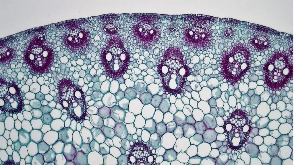
Brightfield Microscope Image

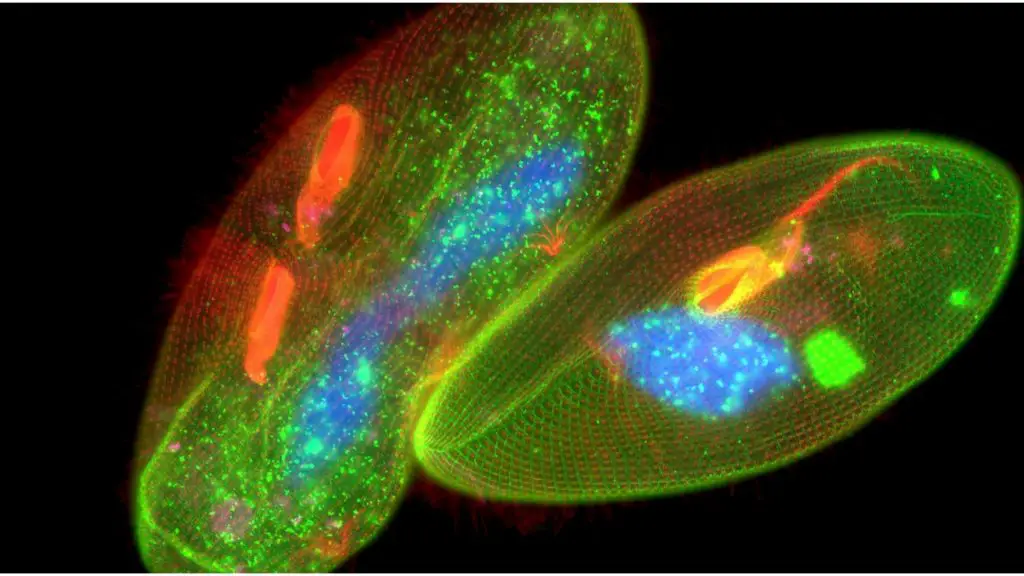
Fluorescence Microscope Image

Reference
- https://microbiologynote.com/fluorescence-microscope-principle-definition-uses-parts/
- https://microbiologynote.com/bright-field-microscope-definition-parts-working-principle-application/
- https://www.news-medical.net/life-sciences/Fluorescence-Microscopy-vs-Light-Microscopy.aspx
- Text Highlighting: Select any text in the post content to highlight it
- Text Annotation: Select text and add comments with annotations
- Comment Management: Edit or delete your own comments
- Highlight Management: Remove your own highlights
How to use: Simply select any text in the post content above, and you'll see annotation options. Login here or create an account to get started.